|
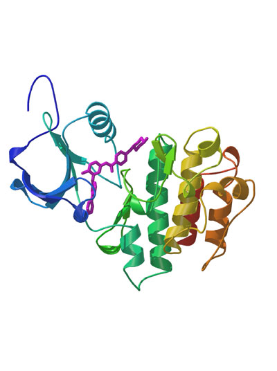
5. Proteomics: the 3-D structure of a cancer-causing protein, BCR-ABL. The protein does not occur naturally but is produced by the fusion of two genes caused by a chromosomal abnormality. The BCR-ABL protein stimulates cell proliferation and may cause a form of leukemia. Shown in purple is a small molecule drug, Gleevec, which inhibits BCR-ABL function. It is with 3-D information like this that drugs will in future be designed to target particular proteins. The computer model of BCR-ABL's structure does not show the details of the atoms or individula amino acids, but nevertheless accurately reflects the protein's lay-out.
Onto the next photo >>
|